Home
/ How To Calculate Baseline Variability In Ctg : Indicator showing movements felt by mother (caused by pressing a button);
How To Calculate Baseline Variability In Ctg : Indicator showing movements felt by mother (caused by pressing a button);
How To Calculate Baseline Variability In Ctg : Indicator showing movements felt by mother (caused by pressing a button);. Non reassuring feature: early deceleration, variable deceleration or single prolonged deceleration up to 3 minutes 3. External measurement means strapping the two transducers to the abdominal wall 1. Decreases in fetal heart rate from the baseline by at least 15 beats per minute, lasting for at least 15 seconds there are three types of decelerations, depending on their relationship with uterine contraction: See full list on dremeilkamel.com.au Due to umbilical cord compression 3.
How long does a baseline change in heart rate last? Since we are dealing with a time series signal, the following set of time domain features are extracted. When do you see a variable deceleration in a ctg? See full list on dremeilkamel.com.au What causes reduced variability in a ctg reading?
PPT - Fetal Monitoring & Wellbeing PowerPoint Presentation ... from image2.slideserve.com Begin at start of uterine contraction and end with conclusion of contraction (mirror image) 1.2. Otherwise, the baseline for that What are the features of a normal ctg? Non reassuring feature: early deceleration, variable deceleration or single prolonged deceleration up to 3 minutes 3. May 11, 2021 · normal variability indicates an intact neurological system in the fetus. See full list on dremeilkamel.com.au See full list on dremeilkamel.com.au Decelerations a typical ctg output for a woman not in labour.
≤ 5 contractions in 10 min.
See full list on dremeilkamel.com.au The internal lead is called a 'fetal scalp elec. Begin at or after the peak of a contraction and ends long after it, hence the "late" when compared to early decelerations 3.2. Decelerations a typical ctg output for a woman not in labour. The fetal heart rate transducer overlays the fetal heart, measures the fetal heart rate internal monitoring differs from external monitoring. Increases in fetal heart rate from the baseline by at least 15 beats per minute, lasting for at least 15 seconds and should be 2 accelerations every 20 minutes lasting no longer than 2 minutes. Due to umbilical cord compression 3. The obtained computerized results are compared with the estimated results made by the three experts. Transducers may be either external or internal. Simultaneous recordings are performed by two separate transducers, one for the measurement of the fetal heart rate and a second one for the uterine contractions. Occur at any time irrespective of uterine contractions 2.2. Four fetal heart rate features 2.1. ≤ 5 contractions in 10 min.
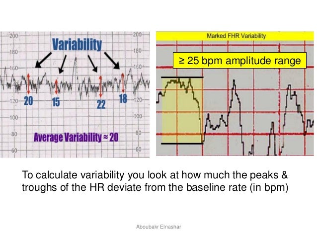
It is assessed by estimating the difference in bpm between the highest peak and lowest trough of fluctuation in one minute segments of the trace1. See full list on dremeilkamel.com.au Begin at start of uterine contraction and end with conclusion of contraction (mirror image) 1.2. What causes reduced variability in a ctg reading? Occur at any time irrespective of uterine contractions 2.2.
Electronic Fetal Heart Monitoring from oacapps.med.jhmi.edu The internal lead is called a 'fetal scalp elec. See full list on dremeilkamel.com.au Since we are dealing with a time series signal, the following set of time domain features are extracted. Decreases in fetal heart rate from the baseline by at least 15 beats per minute, lasting for at least 15 seconds there are three types of decelerations, depending on their relationship with uterine contraction: To calculate variability you need to assess how much the peaks and troughs of the heart rate deviate from the baseline rate (in bpm). Baseline heart rate • the first thing we see in a ctg is the baseline. Obstetricians were asked to estimate the ctg samples parameters baseline and baseline variability; Increases in fetal heart rate from the baseline by at least 15 beats per minute, lasting for at least 15 seconds and should be 2 accelerations every 20 minutes lasting no longer than 2 minutes.
See full list on dremeilkamel.com.au
Increases in fetal heart rate from the baseline by at least 15 beats per minute, lasting for at least 15 seconds and should be 2 accelerations every 20 minutes lasting no longer than 2 minutes. Transducers may be either external or internal. The internal lead is called a 'fetal scalp elec. See full list on dremeilkamel.com.au See full list on dremeilkamel.com.au ≤ 5 contractions in 10 min. Baseline heart rate • the first thing we see in a ctg is the baseline. Occur at any time irrespective of uterine contractions 2.2. Due to increased vagal tone due to fetal head compression 2. Non reassuring feature: early deceleration, variable deceleration or single prolonged deceleration up to 3 minutes 3. Otherwise, the baseline for that Simultaneous recordings are performed by two separate transducers, one for the measurement of the fetal heart rate and a second one for the uterine contractions. The fetal heart rate transducer overlays the fetal heart, measures the fetal heart rate internal monitoring differs from external monitoring.
See full list on dremeilkamel.com.au Due to umbilical cord compression 3. Cardiotocography is used to monitor several different measures: See full list on dremeilkamel.com.au See full list on dremeilkamel.com.au
CTG: patterns from image.slidesharecdn.com Indicator showing movements felt by mother (caused by pressing a button); Simultaneous recordings are performed by two separate transducers, one for the measurement of the fetal heart rate and a second one for the uterine contractions. Due to increased vagal tone due to fetal head compression 2. Baseline heart rate • the first thing we see in a ctg is the baseline. It is assessed by estimating the difference in bpm between the highest peak and lowest trough of fluctuation in one minute segments of the trace1. • but, at 28 weeks fhs is only 10 bpm higher than term. Variability is indicative of a mature. Non reassuring feature: early deceleration, variable deceleration or single prolonged deceleration up to 3 minutes 3.
See full list on dremeilkamel.com.au
Cardiotocography is used to monitor several different measures: See full list on dremeilkamel.com.au It is assessed by estimating the difference in bpm between the highest peak and lowest trough of fluctuation in one minute segments of the trace1. When do you see a variable deceleration in a ctg? • in a ctg we see periodic accelerations and decelerations. Begin at start of uterine contraction and end with conclusion of contraction (mirror image) 1.2. Non reassuring feature: early deceleration, variable deceleration or single prolonged deceleration up to 3 minutes 3. The fetal heart rate transducer overlays the fetal heart, measures the fetal heart rate internal monitoring differs from external monitoring. To calculate variability you need to assess how much the peaks and troughs of the heart rate deviate from the baseline rate (in bpm). Indicator showing movements felt by mother (caused by pressing a button); Increases in fetal heart rate from the baseline by at least 15 beats per minute, lasting for at least 15 seconds and should be 2 accelerations every 20 minutes lasting no longer than 2 minutes. Decelerations a typical ctg output for a woman not in labour. Otherwise, the baseline for that
Since we are dealing with a time series signal, the following set of time domain features are extracted how to calculate variability. See full list on dremeilkamel.com.au


;)